Search the site...

Angeline Mlenar
  • Blog
  • About
  • Contact
  • Blog
  • About
  • Contact

Ways To Diagnose Fallen Arches

7/4/2017

 
Overview

Flat Feet

The most common arch problem is the flat foot. This sometimes starts in childhood or may gradually develop in adulthood. In most cases the flat foot is related to a tight calf. The tightness of the calf forces the foot to overpronate (the inside of the foot rolls inwards) and the arch to break down and collapse. The arch collapse leads to abnormal stress on the plantar fascia leading to heel pain, as well as to the main medial tendon (the posterior tibial tendon), leading to tendonitis and even tears of the tendon. The other common symptom in severe flat feet is pain on the outside of the foot as well as calf and Achilles symptoms.

Causes

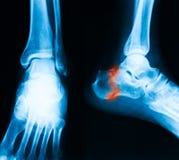
Aging, injury, overuse, or illness can result in fallen arches or a fallen arch on one side. Diabetes. Obesity. Pregnancy. Nerve conditions. Foot abnormalities present since birth. Broken or dislocated bones in the foot. Stretched or torn tendons. Medical conditions such as arthritis. Sudden weight gain

Symptoms

Not everyone who has flat feet experiences symptoms. Others, however, feel persistent pain in their feet and wearing shoes can prove additionally painful. Others only feel symptoms when they walk more than normal, go jogging or participate in a sport that involves running or kicking.

Diagnosis

There are a few simple ways to assess your foot type, and most include making an imprint of your footprint. The classic way is to stand on a hard floor surface with wet feet to make a wet foot print. Look at the narrowest part of your footprint, which should be between your heel and ball of your foot. If the print of your foot in this part is less than 10% of the width of the widest part then you are likely to have high arches. more than 10% but less than 25% then your foot profile is probably normal, more than 25% or even the widest part, then you have flat feet.

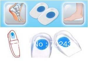
fallen arches support

Non Surgical Treatment

If you have flat feet (pes planus) it does not mean you will have problems or pain. Flat feet are a common condition. Flat feet are defined as an abnormal lowering of the medial longitudinal arch. There is often a hereditary component to the development of flat feet. Flat feet are more common in certain ethnic groups. Flat feet usually occurs equally in both feet and become apparent at an early age. Flat feet can initially be asymptomatic, but can become progressively symptomatic with age. Some flat feet never become bothersome. The medial arch is unstable in flexible flatfoot. The foot can bend as the heel lifts of the ground. This creates stress on the surrounding soft-tissues and joints creating tired, sore arches. The instability of the foot can create abnormal stress that flows up the leg which results in pain in the legs, knees, hips and lower back. Patients with flat feet often present to the chiropodist/podiatrist with problems that are indirectly caused by their foot structure.

Surgical Treatment

Acquired Flat Foot

Procedures may include the following. Fusing foot or ankle bones together (arthrodesis). Removing bones or bony growths, also called spurs (excision). Cutting or changing the shape of the bone (osteotomy). Cleaning the tendons' protective coverings (synovectomy). Adding tendon from other parts of your body to tendons in your foot to help balance the "pull" of the tendons and form an arch (tendon transfer). Grafting bone to your foot to make the arch rise more naturally (lateral column lengthening).

Prevention

Donning a first-rate pair of arch supports, therapeutic socks and proper footwear before heading out to enjoy hours of holiday fun is one option to consider. Your podiatrist can help you find just the right ones. Once you have them on, they?ll help ease the amount of pressure being put on your body and keep the blood flowing in the right direction. While you?re standing in line, consider doing a bit of exercise as well. We?re not talking about channeling your inner Jack LaLanne here. Otherwise, you might attract the attention of the mall security guards. Simple ankle rotations and walking in place may help to reduce edema and give your flat feet a bit of a break. If you happen to be in a shopping mall or center where foot massages are available, take advantage of them periodically. They are likely to make you feel better and it?s a great excuse to carve out a few quiet moments for yourself. If you can?t visit a professional, tuck a personal foot massager into your purse. That way, you can lightly massage your own feet during the car ride home. Lastly, there are certain foods and nutritional supplements available that may reduce edema caused by standing on flat feet for hours at a time. The list includes potassium rich foods like raisins, bananas, baby carrots, nuts and yogurt. So, you may want to pack a snack for those trips to the mall or hit the food court before you hit the stores.

All The Things You Will Need To Understand About

7/2/2017

 
Overview

Pain Under The Heel

Heel pain is a common foot condition. It's usually felt as an intense pain when using the affected heel. Heel pain usually builds up gradually and gets worse over time. The pain is often severe and occurs when you place weight on the heel. In most cases, only one heel is affected, although estimates suggest that around a third of people have pain in both heels. The pain is usually worse first thing in the morning, or when you first take a step after a period of inactivity. Walking usually improves the pain, but it often gets worse again after walking or standing for a long time. Some people may limp or develop an abnormal walking style as they try to avoid placing weight on the affected heel.

Causes

Here are a few in-depth explanations of heel pain causes and potential remedies for those that are afflicted Plantar Fascitis, One of the most well-known causes of heel pain, plantar fascitis occurs when the thick tissue along the arch of the foot becomes tight and inflamed. The foot itself can feel cramped when this tissue is inflamed, causing the afflicted person to feel as if they have a muscle cramp that flexing the foot doesn?t seem to alleviate. Plantar fascitis is most common among men over the age of 40. Warning Signs, The first steps in the morning are extremely painful. Pain flares up after activities like stair or steep hill climbing. Pain continues to occur regardless of the type of shoes or lack of shoes worn.

Symptoms

See your doctor as soon as possible if you experience severe pain accompanied by swelling near your heel. There is numbness or tingling in the heel, as well as pain and fever. There is pain in your heel as well as fever. You are unable to walk normally. You cannot bend your foot downwards. You cannot stand with the backs of the feet raised (you cannot rise onto your toes). You should arrange to see a doctor if the heel pain has persisted for more than one week. There is still heel pain when you are not standing or walking.

Diagnosis

The diagnosis of plantar fasciitis is generally made during the history and physical examination. There are several conditions that can cause heel pain, and plantar fasciitis must be distinguished from these conditions. Pain can be referred to the heel and foot from other areas of the body such as the low back, hip, knee, and/or ankle. Special tests to challenge these areas are performed to help confirm the problem is truly coming from the plantar fascia. An X-ray may be ordered to rule out a stress fracture of the heel bone and to see if a bone spur is present that is large enough to cause problems. Other helpful imaging studies include bone scans, MRI, and ultrasound. Ultrasonographic exam may be favored as it is quick, less expensive, and does not expose you to radiation. Laboratory investigation may be necessary in some cases to rule out a systemic illness causing the heel pain, such as rheumatoid arthritis, Reiter's syndrome, or ankylosing spondylitis. These are diseases that affect the entire body but may show up at first as pain in the heel.

Non Surgical Treatment

Treatment of plantar fasciitis is usually performed in stages according to the duration and degree of pain. Treatment may take many months if the condition has been longstanding. Treatment usually begins with anti-inflammatory medication, shoe modification, temporary limitation of activities, weight loss and heel cord stretching. Also, night splints are often helpful to stretch the plantar fascia. An arch support (orthotic) may also be helpful, especially if you have a flat foot. If the problem continues, the tender area occasionally may be injected with cortisone and a local anesthetic. For a difficult, chronic problem, a period of casting may be used to improve this condition. Surgical treatment is rarely needed. If performed, it aims to partially release the plantar fascia and stimulate healing of the chronic inflammation. Removal of a heel spur, if it is large, may also be done at the time of surgery.

Surgical Treatment

When a diagnosis of plantar fasciitis is made early, most patients respond to conservative treatment and don?t require surgical intervention. Often, when there is a secondary diagnosis contributing to your pain, such as an entrapped nerve, and you are non-responsive to conservative care, surgery may be considered. Dr. Talarico will discuss all options and which approach would be the most beneficial for your condition.

heel spur exercises

Prevention

Feet Pain

The following steps will help prevent plantar fasciitis or help keep the condition from getting worse if you already have it. The primary treatment is rest. Cold packs application to the area for 20 minutes several times a day or after activities give some relief. Over-the-counter pain medications can help manage the pain, consult your healthcare professional. Shoes should be well cushioned, especially in the midsole area, and should have the appropriate arch support. Some will benefit from an orthotic shoe insert, such as a rubber heel pad for cushioning. Orthotics should be used in both shoes, even if only one foot hurts. Going barefoot or wearing slipper puts stress on your feet. Put on supportive shoes as soon as you get out of bed. Calf stretches and stretches using a towel (place the towel under the ball of your feet and pull gently the towel toward you and hold a few seconds) several times a day, especially when first getting up in the morning. Stretching the Achilles tendon at the back of the heel is especially important before sports, but it is helpful for nonathletes as well. Increasing your exercise levels gradually. Staying at a healthy weight. Surgery is very rarely required.

Leg Length Discrepancy Measurements

6/29/2017

 
Overview

Every person?s body is unique and will show different symptoms due to a short leg. Athletes are able to distinguish the negative effects of a leg length that is just 3 mm shorter then the other. A whole host of negative effects can occur to the body that can create chronic pain and may necessitate surgical interventions. The effect of a short leg can be seen almost everywhere in the body.Leg Length Discrepancy

Causes

Some causes of leg length discrepancy (other than anatomical). Dysfunction of the hip joint itself leading to compensatory alterations by the joint and muscles that impact on the joint. Muscle mass itself, i.e., the vastus lateralis muscle, pushes the iliotibial band laterally, causing femoral compensations to maintain a line of progression during the gait cycle. This is often misdiagnosed as I-T band syndrome and subsequently treated incorrectly. The internal rotators of the lower limb are being chronically short or in a state of contracture. According to Cunningham's Manual of Practical Anatomy these are muscles whose insertion is lateral to the long axis of the femur. The external rotators of the hip joint are evidenced in the hip rotation test. The iliosacral joint displays joint fixations on the superior or inferior transverse, or the sagittal axes. This may result from many causes including joint, muscle, osseous or compensatory considerations. Short hamstring muscles, i.e., the long head of the biceps femoris muscle. In the closed kinetic chain an inability of the fibula to drop inferior will result in sacrotuberous ligament loading failure. The sacroiliac joint dysfunctions along its right or left oblique axis. Failure or incorrect loading of the Back Force Transmission System (the longitudinal-muscle-tendon-fascia sling and the oblique dorsal muscle-fascia-tendon sling). See the proceedings of the first and second Interdisciplinary World Congress on Low Back Pain. Sacral dysfunction (nutation or counternutation) on the respiratory axis. When we consider the above mentioned, and other causes, it should be obvious that unless we look at all of the causes of leg length discrepancy/asymmetry then we will most assuredly reach a diagnosis based on historical dogma or ritual rather than applying the rules of current differential diagnosis.

Symptoms

The patient/athlete may present with an altered gait (such as limping) and/or scoliosis and/or low back pain. Lower extremity disorders are possibly associated with LLD, some of these are increased hip pain and degeneration (especially involving the long leg). Increased risk of: knee injury, ITB syndrome, pronation and plantar fascitis, asymmetrical strength in lower extremity. Increased disc or vertebral degeneration. Symptoms vary between patients, some patients may complain of just headaches.

Diagnosis

Limb length discrepancy can be measured by a physician during a physical examination and through X-rays. Usually, the physician measures the level of the hips when the child is standing barefoot. A series of measured wooden blocks may be placed under the short leg until the hips are level. If the physician believes a more precise measurement is needed, he or she may use X-rays. In growing children, a physician may repeat the physical examination and X-rays every six months to a year to see if the limb length discrepancy has increased or remained unchanged. A limb length discrepancy may be detected on a screening examination for curvature of the spine (scoliosis). But limb length discrepancy does not cause scoliosis.

Non Surgical Treatment

You and your physician should discuss whether treatment is necessary. For minor LLDs in adults with no deformity, treatment may not be necessary. Because the risks may outweigh the benefits, surgical treatment to equalize leg lengths is usually not recommended if the difference is less than one inch. For these small differences, your physician may recommend a shoe lift. A lift fitted to the shoe can often improve your walking and running, as well as relieve back pain caused by LLD. Shoe lifts are inexpensive and can be removed if they are not effective. They do, however, add weight and stiffness to the shoe.

Leg Length Discrepancy

what happens if one leg is shorter than the other?

Surgical Treatment

Your child will be given general anesthetic. We cut the bone and insert metal pins above and below the cut. A metal frame is attached to the pins to support the leg. Over weeks and months, the metal device is adjusted to gradually pull the bone apart to create space between the ends of the bones. New bone forms to fill in the space, extending the length of the bone. Once the lengthening process is completed and the bones have healed, your child will require one more short operation to remove the lengthening device. We will see your child regularly to monitor the leg and adjust the metal lengthening device. We may also refer your child to a physical therapist to ensure that he or she stays mobile and has full range of motion in the leg. Typically, it takes a month of healing for every centimeter that the leg is lengthened.

What Is Mortons Neuroma

6/4/2017

 
Overview

intermetatarsal neuromaThis is a painful condition affecting a small nerve in the foot. It occurs when the five long bones that run the length of the foot get pushed together, pinching the nerve in between. This friction on the nerve causes it to thicken and inflame causing pain. The condition gets its name from an American surgeon, George Morton.

Causes

Poorly fitted footwear can be a cause. Shoes that have a tight and narrow toe box can cause the never to become entrapped causing the pain. High heeled shoes abnormally place the metatarsals under extreme pressure which can cause Morton?s Neuroma. In cases of abnormal pronation, there can be significant motion between the 3rd and 4th metatarsals which can cause an irritation to the nerve that runs between them. This inflammation causes the pain.

Symptoms

Patients will complain of numbness, a ?pins and needles? type of tingling and loss of sensation in the toes. Burning pain in the ball of the foot that may radiate into the toes. The pain generally intensifies with activity or wearing shoes. Night pain is rare. There may also be numbness in the toes, or an unpleasant feeling in the toes. Runners may feel pain as they push off from the starting block. High-heeled shoes, which put the foot in a similar position to the push-off, can also aggravate the condition. Tight, narrow shoes also aggravate this condition by compressing the toe bones and pinching the nerve.

Diagnosis

Your health care provider can usually diagnose this problem by examining your foot. A foot x-ray may be done to rule out bone problems. MRI or high-resolution ultrasound can successfully diagnose Morton's neuroma. Nerve testing (electromyography) cannot diagnose Morton's neuroma, but may be used to rule out conditions that cause similar symptoms. Blood tests may be done to check for inflammation-related conditions, including certain forms of arthritis.

Non Surgical Treatment

Initial therapies are nonsurgical and relatively simple. They can involve one or more of the following treatments. Changes in footwear. Avoid high heels or tight shoes, and wear wider shoes with lower heels and a soft sole. This enables the bones to spread out and may reduce pressure on the nerve, giving it time to heal. Orthoses. Custom shoe inserts and pads also help relieve irritation by lifting and separating the bones, reducing the pressure on the nerve. Injection. One or more injections of a corticosteroid medication can reduce the swelling and inflammation of the nerve, bringing some relief. Several studies have shown that a combination of roomier, more comfortable shoes, nonsteroidal anti-inflammatory medication, custom foot orthoses and cortisone injections provide relief in over 80 percent of people with Morton's Neuroma.interdigital neuroma

Surgical Treatment

Majority of publications including peer review journal articles, surgical technique description and textbooks promote surgical excision as a gold standard treatment. Surgical excision is described as the most definitive mode of treatment for symptomatic Morton?s neuroma with reported success rates varying between 79% and 93%. Various surgical techniques are described, essentially categorised as dorsal versus plantar incision approaches. Beyond this the commonest technical variation described as influencing the outcome of surgery involves burying and anchoring transacted nerve into soft tissue such as muscle.

The Ideal Solution To Leg Length Discrepancy Are Shoe Lifts

2/19/2016

 
There are actually two different types of leg length discrepancies, congenital and acquired. Congenital means that you are born with it. One leg is anatomically shorter compared to the other. As a result of developmental stages of aging, the human brain senses the walking pattern and identifies some variance. The human body usually adapts by tilting one shoulder to the "short" side. A difference of under a quarter inch isn't grossly excessive, does not need Shoe Lifts to compensate and ordinarily won't have a profound effect over a lifetime.

Shoe Lift

Leg length inequality goes largely undiscovered on a daily basis, however this problem is very easily fixed, and can eliminate numerous cases of back ache.

Treatment for leg length inequality typically consists of Shoe Lifts. They are low cost, commonly costing less than twenty dollars, compared to a custom orthotic of $200 or maybe more. Differences over a quarter inch can take their toll on the spine and should probably be compensated for with a heel lift. In some cases, the shortage can be so extreme that it requires a full lift to both the heel and sole of the shoe.

Chronic back pain is easily the most prevalent ailment impacting people today. Over 80 million men and women suffer from back pain at some stage in their life. It is a problem which costs employers millions of dollars every year on account of lost time and output. New and superior treatment solutions are always sought after in the hope of lowering economic impact this condition causes.

Leg Length Discrepancy Shoe Lifts

People from all corners of the world suffer the pain of foot ache as a result of leg length discrepancy. In a lot of these situations Shoe Lifts can be of very beneficial. The lifts are capable of decreasing any discomfort in the feet. Shoe Lifts are recommended by countless professional orthopaedic practitioners".

So that they can support the human body in a nicely balanced fashion, your feet have got a significant role to play. Despite that, it is often the most neglected region in the human body. Some people have flat-feet meaning there may be unequal force exerted on the feet. This will cause other areas of the body including knees, ankles and backs to be impacted too. Shoe Lifts guarantee that correct posture and balance are restored.

Does A Posterior Calcaneal Spur Hurt?

9/29/2015

 
Calcaneal Spur

Overview

A heel spur is a pointed bony outgrowth of the bone of the heel (the calcaneus bone). Heel spurs under the sole of the foot (plantar area) are associated with plantar fasciitis. Heel spurs and plantar fasciitis can occur alone or be related to underlying diseases. Heel spurs and plantar fasciitis are treated by measures that decrease the associated inflammation and avoid reinjury.

Causes

A strong band of sinew (plantar fascia) stretches across the sole of the foot below the surface of the skin and is attached to a point in the middle of the under surface of the heel bone. With repeated activity on our feet, the plantar fascia can become tight and cause persistent traction (tugging) on its attachment point into the heel bone, and inflammation and pain may develop at this site. This painful condition is known as plantar fasciitis. Sometimes a ?spur? develops at the site of this traction on the bone and protrudes into the surrounding tissue. This is a heel spur.

Posterior Calcaneal Spur

Symptoms

Heel spurs are most noticeable in the morning when stepping out of bed. It can be described as sharp isolated pain directly below the heel. If left untreated heel spurs can grow and become problematic long-term.

Diagnosis

Most patients who are suffering with heel spurs can see them with an X-ray scan. They are normally hooked and extend into the heel. Some people who have heel spur may not even have noticeable symptoms, although could still be able to see a spur in an X-ray scan.

Non Surgical Treatment

Heel spurs are considered a self-limited condition, which means that by making small alterations in your lifestyle and regular routines you can often control the condition. The goal is to relieve pain, reduce friction and transfer pressure from your sensitive foot areas. By eliminating the cause of the heel spur and plantar fasciitis (i.e. better shoes, orthotics to fix your gait, losing weight) will help reduce the pressure put on your fascia and heel and can reduce the inflammation caused by your heel spur. Failure to see improvements after conservative treatments may make surgery your only option.

Surgical Treatment

More than 90 percent of people get better with nonsurgical treatments. If conservative treatment fails to treat symptoms of heel spurs after a period of 9 to 12 months, surgery may be necessary to relieve pain and restore mobility. Surgical techniques include release of the plantar fascia, removal of a spur. Pre-surgical tests or exams are required to identify optimal candidates, and it's important to observe post-surgical recommendations concerning rest, ice, compression, elevation of the foot, and when to place weight on the operated foot. In some cases, it may be necessary for patients to use bandages, splints, casts, surgical shoes, crutches, or canes after surgery. Possible complications of heel surgery include nerve pain, recurrent heel pain, permanent numbness of the area, infection, and scarring. In addition, with plantar fascia release, there is risk of instability, foot cramps, stress fracture, and tendinitis.

Prevention

Heel Spur symptoms can be prevented from returning by wearing proper shoes and using customized orthotics and insoles to relieve pressure. It is important to perform your exercises to help keep your foot stretched and relaxed.

What May Cause Calcaneal Spur

9/22/2015

 
Inferior Calcaneal Spur

Overview

A heel spur occurs when calcium deposits build up on the underside of the heel bone. The abnormal calcium deposits form when the plantar fascia pulls away from the heel. This stretching of the plantar fascia is common among people who have flat feet, but people with unusually high arches can also develop this problem. Heel spurs are especially common among athletes who do a lot of running and jumping. Also, women who wear high heels have a significantly higher incidence of heel spurs. Still, it can happen to anyone.

Causes

A heel spur usually develops as a result of wear and tear over time, which leads to the degeneration of connective tissue called fascia. Standing for prolonged periods and wearing shoes that do not provide the right type of arch support can also lead to connective tissue damage in the heel. The body attempts to repair the damaged tissue by delivering calcium to the affected region, but sometimes too much calcium begins to accumulate and this results in painful plantar fasciitis.

Inferior Calcaneal Spur

Symptoms

Most of the time heel spurs present as pain in the region surrounding the spur, which typically increases in intensity after prolonged periods of rest. Patients may not be able to bear weight on the afflicted heel comfortably. Running, walking, or lifting heavy weight may exacerbate the issue.

Diagnosis

A heel spur is often seen on X-ray as a bony protrusion, which can vary in size. However, because a Heel Spur only indicates increased load on the plantar fascia, and not pain, an ultra sound may be required to assess other actual cause of the heel pain such and may include checking to see if the plantar fascia is inflamed or degenerated.

Non Surgical Treatment

Heel spurs and plantar fasciitis are treated by measures that decrease the associated inflammation and avoid reinjury. Local ice applications both reduce pain and inflammation. Physical therapy methods, including stretching exercises, are used to treat and prevent plantar fasciitis. Anti-inflammatory medications, such as ibuprofen or injections of cortisone, are often helpful. Orthotic devices or shoe inserts are used to take pressure off plantar spurs (donut-shaped insert), and heel lifts can reduce stress on the Achilles tendon to relieve painful spurs at the back of the heel. Similarly, sports running shoes with soft, cushioned soles can be helpful in reducing irritation of inflamed tissues from both plantar fasciitis and heel spurs. Infrequently, surgery is performed on chronically inflamed spurs.

Surgical Treatment

When chronic heel pain fails to respond to conservative treatment, surgical treatment may be necessary. Heel surgery can provide pain relief and restore mobility. The type of procedure used is based on examination and usually consists of releasing the excessive tightness of the plantar fascia, called a plantar fascia release. The procedure may also include removal of heel spurs.

What Is Bursitis Of The Foot?

8/25/2015

 
Overview

A bursa is a small sack of fluid that sits between a tendon and a bone to help the tendon move smoothly over the bone. The retrocalcaneal bursa in situated in the feet between the Achilles tendon and the calcaneus or heel bone. With repeated trauma the bursa can become inflamed. Achilles tendon bursitis is often mistaken for achilles tendinitis. It is possible for the athlete to have both achilles tendinitis and achilles tendon bursitis at the same time which is known as Haglund's syndrome.

Causes

A bursa acts as a cushion and lubricant between tendons or muscles sliding over bone. There are bursas around most large joints in the body, including the ankle. The retrocalcaneal bursa is located in the back of the ankle by the heel. It is where the large Achilles tendon connects the calf muscles to the heel bone. Repeated or too much use of the ankle can cause this bursa to become irritated and inflamed. Possible causes are too much walking, running, or jumping. This condition is usually linked to Achilles tendinitis. Sometimes retrocalcaneal bursitis may be mistaken for Achilles tendinitis. Risks for this condition include starting an aggressive workout schedule, or suddenly increasing activity level without the right conditioning.

Symptoms

When the bursa becomes inflamed after an injury, symptoms usually develop suddenly. When the bursa develops without an injury, symptoms may develop gradually. With both posterior and anterior Achilles tendon bursitis, symptoms usually include swelling and warmth at the back of the heel. A minimally red, swollen, tender spot develops on the back of the heel. When the inflamed bursa enlarges, it appears as a red lump under the skin of the heel and causes pain at and above the heel. If posterior Achilles tendon bursitis becomes chronic, the swelling may become hard, fluid-filled, and red or flesh-colored.

Diagnosis

A good clinical practise includes evaluation of the tendon, bursa and calcaneum by, careful history, inspection of the region for bony prominence and local swelling as well as palpation of the area of maximal tenderness. Biomechanical abnormalities, joint stiffness and proximal soft tissue tightening can exacerbate an anatomical predisposition to retrocalcaneal bursitis, they warrant correction when present.

Non Surgical Treatment

Most patients with achilles and retrocalcaneal bursitis heal well with appropriate physiotherapy and other usual bursitis conventional and natural cures being administered. Specific treatments for ankle / heel bursitis may include footwear correction. Get well-fitting, soft-backed (or even open-backed whenever possible) shoes for both day to day wear and exercise. High-heels should really be a no no or worn sparingly, ladies. Heel protectors. Heel pads and heel lifts are great simple solutions to cushion and protect the Achilles area from the irritation of the shoes. Orthotics. There are various orthotic devices out there (some only available over-the-counter). One example is a custom arch suppport. These can control abnormal motion in your feet by lining them up correctly in your shoes to help you move in the right matter so the bursitis heals faster and does not return back again. Exercise modification Stretch your heel, mainly Achilles tendon, frequently, particularly before and after excercise or prolonged sitting. If you are a jogger, try to run on softer surfaces (no hard concrete, please). Running uphill training is best to be avoided by Achilles and retrocalcaneal bursitis sufferers.

Surgical Treatment

Surgery is rarely need to treat most of these conditions. A patient with a soft tissue rheumatic syndrome may need surgery, however, if problems persist and other treatment methods do not help symptoms.

Pain After Hammertoe Surgery

6/26/2015

 
Hammer ToeOverview

A Hammer toe is a toe that tends to remain bent at the middle joint in a claw-like position. There are 2 types of hammer toe. Flexible hammer toe, can be straightened by hand. Rigid hammer toe, cannot be pulled straight and can hammertoes be extremely painful. The position of the toe can also lead to corns or calluses. These may also be painful. Hammer toe may be present at birth or develop later in life due to tendons that have tightened, causing the toe's joints to curl downward. Occasionally, all toes may be bent. This may be due to problems with the peripheral nerves or the spinal cord.

Causes

Medical problems, such as stroke or diabetes that affect the nerves, may also lead to hammertoe. For example, diabetes can result in poor circulation, especially in the feet. As a result, the person may not feel that their toes are bent into unnatural positions. The likelihood of developing hammertoe increases with age and may be affected by gender (more common in women) and toe length; for example, when the second toe is longer than the big toe, hammertoe is more likely to occur. Hammertoe may also be present at birth. Genetics may factor in to developing hammertoe, particularly if the foot is flat or has a high arch, resulting in instability.

Hammer ToeSymptoms

A toe (usually the second digit, next to the big toe) bent at the middle joint and clenched into a painful, clawlike position. As the toe points downward, the middle joint may protrude upward. A toe with an end joint that curls under itself. Painful calluses or corns. Redness or a painful corn on top of the bent joint or at the tip of the affected toe, because of persistent rubbing against shoes Pain in the toes that interferes with walking, jogging, dancing, and other normal activities, possibly leading to gait changes.

Diagnosis

Some questions your doctor may ask of you include, when did you first begin having foot problems? How much pain are your feet or toes causing you? Where is the pain located? What, if anything, seems to improve your symptoms? What, if anything, appears to worsen your symptoms? What kind of shoes do you normally wear? Your doctor can diagnose hammertoe or mallet toe by examining your foot. Your doctor may also order X-rays to further evaluate the bones and joints of your feet and toes.

Non Surgical Treatment

If the problem is caught in the early stages you can avoid hammer toe surgery. One of the easiest methods of treatment is to manipulate the toe out of a bent position then splint and buddy wrap it alongside it?s larger neighbour. This method of hammer toe taping will help the problem to fix itself. Make sure the toe isn?t resuming its bent shape during the recovery. To alleviate some of the painful symptoms of hammer toe avoid wearing high heels or shoes that cramp or stifle your feet. Choosing a pair of minimalist shoes can be an excellent choice for both foot and postural health. Wearing shoes that give the toes plenty of space and are comfortable lined is also a smart choice. Hammer toe recovery starts be treating the toe respectfully. Soft insoles or protection for the corn can also provide additional assistance.

Surgical Treatment

Joint resection procedures involves removing part of one of the two small joints of the toe directly underneath where the digit is crooked. The purpose is to make room for the toe to be re-positioned flat or straight. Because hammer toes become rigid or fixed with time, removing the joint becomes the only option when the knuckle is stiff. Its important to understand that this procedure does not involve the joint of the ball of the foot, rather the a small joint of the toe. Medical terminology for this procedure is called a proximal interphalangeal joint arthroplasty or a distal interphalangeal joint arthroplasty, with the latter involving the joint closer to the tip of the toe.

HammertoePrevention

How can I prevent hammer toe? Avoid wearing shoes that are narrow or don?t fit well. Also, don?t wear heels higher than 2 inches. Instead, choose shoes with a wide toe box that give you ? inch between the end of your longest toe and the inside tip of the shoe. Check often to make sure your child?s shoes fit, especially when he or she is having a growth spurt.

Hammertoe Pain

6/25/2015

 
HammertoeOverview

Hammer toes a bending and hardening of the joints of the second, third, fourth, or fifth toes. If you look down at your feet and you can?t see the tips of the toenails, you might suffer from hammertoe. Early signs of hammertoe are a bend in the joint of any toe except the big toe. The bend in the joint causes the top of the toe to appear to curl under as if it?s ?hammering? into the floor.

Causes

Hammer toe is commonly caused by wearing shoes that are too narrow, tight or short on a regular basis. By doing so, your toe joints are forced into odd position. Over time, the tendons and muscles in your toe become shorter and cause it to bend. You can suffer a hammer toe if you have diabetes and the disease is worsening. If this occurs, you should contact your doctor right away. Arthritis can also cause hammer toes. Because your toe muscles get out of balance when you suffer from this joint disorder, tendons and joints of your toes are going to experience a lot of pressure.

HammertoeSymptoms

The most obvious symptoms of this injury will be the the middle toe joint is permanently bent at an angle. In the beginning movement may still be possible but as time passes and the injury worsens the toe will be locked in place and possible require hammer toe correction surgery to fix. Another key indicator of hammer toe is that a lump or corn will form on top of the toe. The toe joint will be painful and walking can Hammer toe cause severe discomfort. Occasionally a callus may form on the sole of the injured foot. If you see any of these symptoms together or have been enduring pain for some time, seeing a podiatrist should be your next step.

Diagnosis

Your healthcare provider will examine your foot, checking for redness, swelling, corns, and calluses. Your provider will also measure the flexibility of your toes and test how much feeling you have in your toes. You may have blood tests to check for arthritis, diabetes, and infection.

Non Surgical Treatment

Orthotics are shoe inserts that can help correct mechanical foot-motion problems to correct pressure on your toe or toes and reduce pain. Changing shoes. You should seek out shoes that conform to the shape of your feet as much as possible and provide plenty of room in the toe box, ensuring that your toes are not pinched or squeezed. You should make sure that, while standing, there is a half inch of space for your longest toe at the end of each shoe. Make sure the ball of your foot fits comfortably in the widest part of the shoe. Feet normally swell during the course of the day, so shop for shoes at the end of the day, when your feet are at their largest. Don't be vain about your shoe size, sizes vary by brand, so concentrate on making certain your shoes are comfortable. Remember that your two feet are very likely to be different sizes and fit your shoe size to the larger foot. Low-heel shoes. High heels shift all your body weight onto your toes, tremendously increasing the pressure on them and the joints associated with them. Instead, wear shoes with low (less than two inches) or flat heels that fit your foot comfortably.

Surgical Treatment

If conservative treatments fail and your symptoms persist, the doctor may recommend a surgical option to straighten the toe. The procedures used vary greatly, depending upon the reasons for the hammertoe. There are a number of different operations to correct hammertoes, the most common ones involve Soft tissue corrections such as tendon transfers, tendon lengthening, and joint capsule repairs. Digital arthroplasty involves removal of bone from the bent joint to allow the toe to straighten. The temporary use of pins or K-wires may be necessary to keep the toe straight during the healing period. Joint implants are sometimes used to allow for a better range of motion in the toe following surgery. Digital arthrodesis involves the removal of bone from the bent joint and fusing the toe in a straight position. If the corn is due to a bone spur, the most common procedure used is an exostectomy, in which surgically removing it or filing it down removes the bone spur. Because of the possible complications involved with any surgery, one should be sure to understand the risks that may be involved with surgery to correct hammertoes and remove bone spurs.
<<Previous
Powered by Create your own unique website with customizable templates.